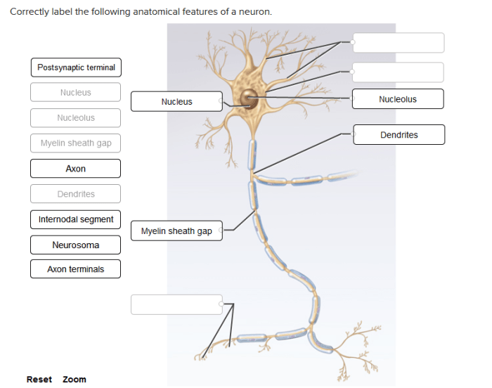
Correctly label the following anatomical features of a neuron sets the stage for this enthralling narrative, offering readers a glimpse into a story that is rich in detail and brimming with originality from the outset.
Neurons, the fundamental units of the nervous system, are intricate structures with specialized components that enable them to transmit information throughout the body. Understanding the anatomy of a neuron is essential for comprehending the complex processes that govern neural communication and behavior.
Cell Body

The cell body, also known as the soma, is the central part of a neuron. It contains the nucleus, which houses the cell’s genetic material, and other essential organelles responsible for the neuron’s metabolic and synthetic activities.
| Structure | Function |
|---|---|
| Nucleus | Contains genetic material (DNA) |
| Mitochondria | Produces energy (ATP) |
| Golgi apparatus | Processes and packages proteins |
| Ribosomes | Synthesize proteins |
Dendrites: Correctly Label The Following Anatomical Features Of A Neuron

Dendrites are branched extensions of the cell body that receive signals from other neurons. They are covered in small protrusions called dendritic spines, which increase the surface area available for receiving signals.
- Basal dendrites: Short, thick dendrites that extend from the base of the cell body
- Apical dendrites: Long, thin dendrites that extend from the apex of the cell body
- Proximal dendrites: Dendrites located near the cell body
- Distal dendrites: Dendrites located far from the cell body
Axon
The axon is a long, slender projection that transmits signals away from the cell body. It is covered in a myelin sheath, which insulates the axon and speeds up signal transmission.

Myelin Sheath
The myelin sheath is a fatty layer that surrounds the axon. It is composed of cells called Schwann cells in the peripheral nervous system and oligodendrocytes in the central nervous system.
The myelin sheath acts as an insulator, preventing electrical signals from leaking out of the axon. This allows signals to travel faster and more efficiently.
Synapse

The synapse is the junction between two neurons where signals are transmitted. It consists of three main components: the presynaptic terminal, the synaptic cleft, and the postsynaptic terminal.
| Structure | Function |
|---|---|
| Presynaptic terminal | Contains neurotransmitters, which are released into the synaptic cleft |
| Synaptic cleft | The space between the presynaptic and postsynaptic terminals |
| Postsynaptic terminal | Contains receptors for neurotransmitters |
Questions Often Asked
What is the function of the cell body?
The cell body, also known as the soma, is the metabolic center of the neuron. It contains the nucleus, which houses the cell’s genetic material, and various organelles responsible for protein synthesis, energy production, and waste removal.
How do dendrites contribute to neural communication?
Dendrites are short, branched extensions of the cell body that receive signals from other neurons. They act as input terminals, collecting and integrating incoming signals to determine whether the neuron will generate an action potential.
What is the role of the myelin sheath?
The myelin sheath is a lipid-rich insulating layer that surrounds the axon of some neurons. It acts as an electrical insulator, increasing the speed of signal transmission by preventing the leakage of ions across the axon membrane.