Compound dysplastic nevus with mild atypia positive margins, a peculiar skin lesion, has garnered attention in the medical community. This article delves into its clinical presentation, histopathology, differential diagnosis, management, and prognosis, providing a comprehensive overview of this intriguing condition.
These lesions, characterized by their distinctive clinical features and microscopic findings, pose diagnostic challenges and warrant careful evaluation to guide appropriate treatment and follow-up.
Compound Dysplastic Nevus with Mild Atypia Positive Margins

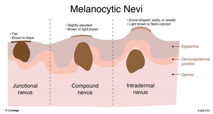
Compound dysplastic nevus with mild atypia positive margins is a type of atypical nevus that exhibits features of both compound nevi and dysplastic nevi. It is characterized by a compound architectural pattern, mild cellular atypia, and positive surgical margins.
Terminology and Definition
Compound dysplastic nevi are melanocytic lesions that demonstrate a dual architectural pattern, with both intradermal and epidermal components. Dysplastic nevi, on the other hand, exhibit atypical cellular features, such as increased nuclear size and pleomorphism. Compound dysplastic nevi with mild atypia positive margins combine these characteristics, presenting with a compound architectural pattern and mild cellular atypia, as well as positive margins indicating incomplete surgical excision.
Clinical Presentation
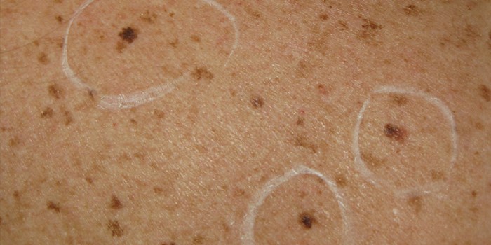
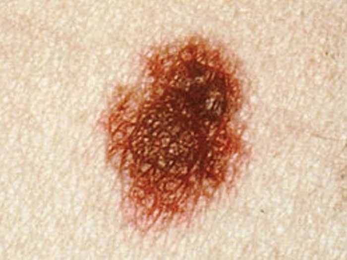
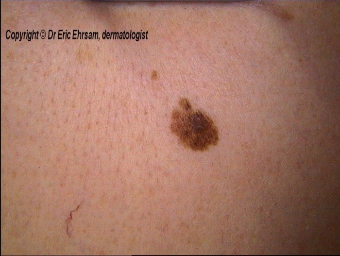
Compound dysplastic nevi with mild atypia typically present as slightly elevated, round or oval lesions ranging in size from a few millimeters to several centimeters. They often have irregular borders and a variegated color, with shades of tan, brown, or pink.
These lesions can occur anywhere on the skin, but are most commonly found on sun-exposed areas, such as the face, arms, and legs.
Histopathology, Compound dysplastic nevus with mild atypia positive margins
Histopathologically, compound dysplastic nevi with mild atypia demonstrate a compound architectural pattern, with both intradermal and epidermal components. The intradermal component consists of nests and cords of melanocytes arranged in a haphazard fashion. The epidermal component exhibits mild cellular atypia, including increased nuclear size and pleomorphism, as well as occasional mitotic figures.
Differential Diagnosis
The differential diagnosis of compound dysplastic nevi with mild atypia positive margins includes other atypical nevi, such as dysplastic nevi and Spitz nevi. Dysplastic nevi typically exhibit more severe cellular atypia and a more complex architectural pattern, while Spitz nevi are characterized by prominent nucleoli and a pushing border.
Other lesions that may need to be considered in the differential diagnosis include melanoma in situ and superficial spreading melanoma.
Management and Treatment
The recommended management for compound dysplastic nevi with mild atypia positive margins is complete surgical excision with clear margins. This involves removing the lesion and a surrounding margin of normal skin to ensure complete removal of all atypical cells. In some cases, additional treatment, such as Mohs micrographic surgery or radiation therapy, may be necessary to achieve clear margins.
Prognosis and Follow-up
The prognosis for patients with compound dysplastic nevi with mild atypia positive margins is generally good. However, these lesions have an increased risk of developing into melanoma compared to typical nevi. Therefore, regular follow-up examinations are recommended to monitor for any changes in the lesion or the development of new lesions.
Frequently Asked Questions
What is the significance of positive margins in compound dysplastic nevus?
Positive margins indicate the presence of dysplastic cells at the edge of the excised tissue, suggesting incomplete removal. This necessitates further excision to ensure complete clearance and reduce the risk of recurrence.
How does compound dysplastic nevus differ from other types of dysplastic nevi?
Compound dysplastic nevus involves both the epidermis and the dermis, while other types of dysplastic nevi are confined to the epidermis. This distinction is important for diagnosis and management, as compound nevi have a higher risk of malignant transformation.
What are the potential complications associated with compound dysplastic nevus?
The primary concern is the potential for malignant transformation into melanoma. Other complications include inflammation, bleeding, and scarring.


